Plant Imaging Unit

Research Interests
We develop new assays and analysis pipelines for plant biology using our expertise in various microscopy fields: histology, fluorescence microscopy, transmission and scanning electron microscopy and image analysis. We apply our skills to all the model systems studied in the Department of Plant Sciences (Arabidopsis seeds, seedlings and adult plants, Marchantia, mosses, tobacco and rice, protoplasts, algae, etc.).
Skills
|
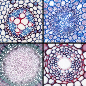
Histology |
Topographic and histochemical colorations on fresh vegetal material (free-hand or vibratome sections) or fixed and included material (microtome slices) using paraffin, Technovit 7100 and Technovit 8100 as embedding media. |
|
|---|---|---|
|
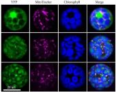
Fluorescence microscopy
|
Use of widefield and confocal imaging for the observation of fluorescent proteins or markers in cells in culture (protoplasts, unicellular algae) or in whole plants, with and without clearing. Immunofluorescence stainings on Arabidopsis seedlings and unicellular algae (done manually, or using the Intavis InsituPro VSi robot).
|
|
|
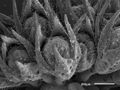
Electron microscopy
|
Transmission electron microscopy for the visualisation of the ultrastructure of plant samples. Scanning electron microscopy to observe surface structures on various types of plants, on fresh material or on fixed organs treated with critical point drying.
|
|
|
Image treatment and analysis
|
Design and implementation of image analysis pipelines using ImageJ/Fiji (and writing of Fiji macros in particular). |
|
We offer our skills in microscopy at the steps of:
- conception and planning of experiments;
- preparation of samples (in particular fixation, embedding, sectioning and staining);
- image acquisition;
- treatment and analysis of pictures.
These services are offered as collaborations with the different laboratories of the Department of Plant Sciences.
Another of our objectives is to develop novel approaches and methods for plant imaging.
In addition, we train users on various kinds of microscopes, and maintain the microscopy equipment of the Department of Plant Sciences (widefield microscopes and stereomicroscopes).
Teaching
Course 12B013: Plant biology
Mandatory course for 2nd-year bachelor students in biology.
This part of the course consists in 12h of lecture and 3 afternoons of practicals dedicated to the study and the observation of the different vegetal tissues and organs.
Course 12B023: Biochemistry – methods (image analysis part)
Mandatory course for 2nd-year bachelor students in biology.
This part of the course consists in 3h of lecture and one practical session dedicated to image treatment and analysis in biology.
Course 14B640: Histology and tissue imaging
Optional course for 3rd year bachelor and master students.
Theoretical and practical course covering classical histology techniques, as well as confocal imaging and image analysis (40h in total).
Inter-university course: Plant imaging: advanced light microscopy
2-days intensive course for PhD students.
Lectures and hands-on sessions covering the preparation and the observation of plant samples by optical microscopy, as well as basic image analysis.
Inter-university course: Image treatment and analysis in microscopy
1.5-day intensive course for PhD students.
Lectures and exercises sessions covering basic as well as more advanced image treatment and analysis.
Course 14B063A: Microscopy and imaging
Optional course for Master students.
Lectures and hands-on sessions covering basic and advanced fluorescence imaging.
Course 14B044: Electron microscopy
Optional course for Master and PhD students and post-docs.
Lectures and hands-on session covering the preparation and the observation of samples by transmission and scanning electron microscopy.
Supervision of Bachelor monographies.
Key Publications
Pesquera M., Martinez J., Maillot B., Wang K., Hofmann M., Raia P., Loubéry S., Steensma P., Hothorn M. & Fitzpatrick T.B.:
Structural and functional studies of Arabidopsis thaliana triphosphate tunnel metalloenzymes reveal roles for additional domains.
J Biol Chem 2022, 298(11):102438. PMID: 36049521. DOI: 10.1016/j.jbc.2022.102438.
De Giorgi J., Fuchs C., Iwasaki M., Kim W., Piskurewicz U., Gully K., Utz-Pugin A., Mène-Saffrané L., Waridel P., Nawrath C., Longoni F.P., Fujita S., Loubéry S. & Lopez-Molina L.:
The Arabidopsis mature endosperm promotes seedling cuticle formation via release of sulfated peptides.
Dev Cell 2021, 56(22):3066-3081.e5. PMID: 34706263. DOI: 10.1016/j.devcel.2021.10.005.
Demonsais L., Utz-Pugin A., Loubéry S. & Lopez-Molina L.:
Identification of tannic cell walls at the outer surface of the endosperm upon Arabidopsis seed coat rupture.
Plant J 2020, 104(3):567-580. PMID: 32985026. DOI: 10.1111/tpj.14994.
Zhu J., Loubéry S., Broger L., Zhang Y., Lorenzo-Orts L., Utz-Pugin A., Fernie A.R., Young-Tae C. & Hothorn M.:
A genetically validated approach for detecting inorganic polyphosphates in plants.
Plant J 2020, 102(3):507-516. PMID: 31816134. DOI: 10.1111/tpj.14642.
Dell'Aglio E., Dalvit I., Loubéry S. & Fitzpatrick T.B.:
Clarification of the dispensability of PDX1.2 for Arabidopsis viability using CRISPR/Cas9.
BMC Plant Biol 2019, 19(1):464. PMID: 31684863. DOI: 10.1186/s12870-019-2071-9.
Lorenzo-Orts L., Witthoeft J., Deforges J., Martinez J., Loubéry S., Placzek A., Poirier Y., Hothorn L.A., Jaillais Y. & Hothorn M.:
Concerted expression of a cell cycle regulator and a metabolic enzyme from a bicistronic transcript in plants.
Nat Plants 2019, 5(2):184-193. PMID: 30737513. DOI: 10.1038/s41477-019-0358-3.
Loubéry S., De Giorgi J., Utz-Pugin A., Demonsais L. & Lopez-Molina L.:
A Maternally Deposited Endosperm Cuticle Contributes to the Physiological Defects of transparent testa Seeds.
Plant Physiol 2018, 177(3):1218-1233. PMID: 29848749. DOI: 10.1104/pp.18.00416.
Yin R., Skvortsova M.Y., Loubéry S. & Ulm R.:
COP1 is required for UV-B-induced nuclear accumulation of the UVR8 photoreceptor.
Proc Natl Acad Sci U S A 2016, 113(30):E4415-22. PMID: 27407149. DOI: 10.1073/pnas.1607074113.